Chemical engineer with a specialty in stem cell biology and automation. I enjoy creating scalable, automated workflows to gather and analyze data which enable data-driven decisions. I typically can be found tinkering with some budget consumer tech (which has come a long way in the last decade) and making it work for biology! I am interested in building solutions for hard problems from the ground up, computer vision for microscopy, and whatever happens when you point a camera at cells long enough.
If you're working on something hard and impactful at the intersection of biology and engineering — let's chat!
Hover any card for a quick look. Click to go deeper.
Active
Validation of Fluent simulations, powered by PTV. Thousands of parameter sets were modelled using PyFluent, modelled with XGBoost, and validated against real data.
Active
Real-time YOLO inference on a live microscope feed — no specialty consumables required. Image aquisition, live cell identification, manual annotation, CSV export.
Early stage
A feature to be used in a lab-assistant robot to help researchers focus on the important tasks
PhD research, extracurriculars, and side projects that shaped how I think. Click into each card to learn more.
Bay Area Lab Automators Hackathon F2025 · San Francisco
Over a single weekend, our team generated and demonstrated a workflow that would allow cell colony identification, robotic handling, and cell picking using an OpenTrons FLEX
Mission Barns · San Francisco
Architected, built, and programmed a LabVIEW-based process control system which enabled richer real-time process tracking, improved consistency, and reduced run failure rate.
PhD research · McGill University
Automated, end-to-end pipeline for live cell imaging, segmentation, and lineage tracking of differentiating stem cells. Large-scale analysis revealed heterogeneity patterns invisible to endpoint assay.
PhD research · McGill University
Micropatterning is a high-resolution technique requiring specialized equipment, long lead time, and extensive user training. Adapted an off-the-shelf craft cutter to bypass these issues and rapidly iterate.
Personal project
Building large adherent bioreactors needs lots of data. Combined an off-the-shelf laser scanner, colorimetric cell staining, and ML-based segmentation methods to increase the amount and quality of data available for informed decisions about bioreactor design.
Master's research · McGill University
Designed and built a microfluidic strain system to apply developmentally relevent strain during cell differentiation
I'm a scientiet/engineer with a specialty in stem cell biology and automation, currently based in San Francisco. I did my undergrad in Chemical Engineering at the University of Waterloo, where I rotated between different internships every term, really giving me the confidence to try new things and "fail fast". My background spans many things: iPSC differentiation, microfabrication, CFD, prototyping, automation and live-cell imaging — but the thread connecting everything is building scalable, automated workflows that turn raw experimental data into decisions.
I'm passionate about problems that can make a true impact on humanity, in healthcare, the environment, or food. I'm especially drawn to problems where you can point a camera at biology long enough and let the data surprise you — computer vision for microscopy, virtual staining on low-cost hardware, high-throughput organoid production for developmental biology and drug screening.
If you're working on something hard at the intersection of biology and engineering — or if you're hiring — reach out.
When we simulate a system, how do we know the model is correct — especially when we can't see inside a typical stainless steel bioreactor?
CFD simulations of bioreactor internals depend on input parameters that are rarely measured directly — they're guessed or borrowed from literature for different geometries. Without ground-truth flow data, there's no way to close the loop between simulation and reality.
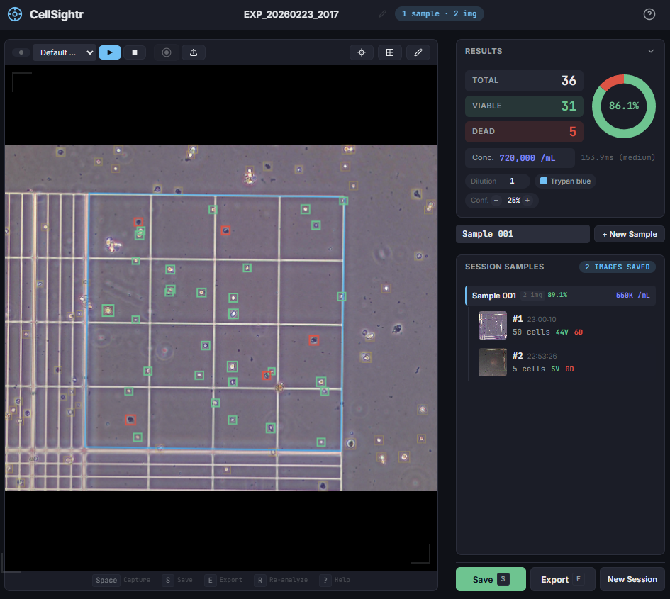
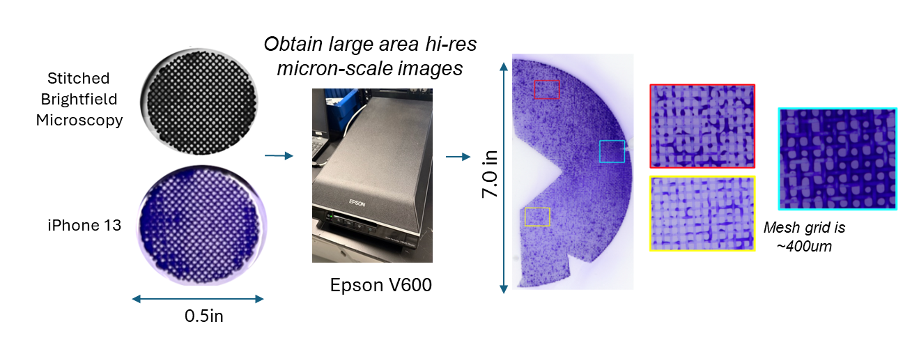
To get that ground truth, I led a team to design fully transparent bioreactor analogs that allow optical access to the flow field. We seeded the fluid with fluorescent tracer beads, illuminated a single plane, and captured footage using consumer phone cameras — no expensive laser-based PIV rigs required. From there, we built a video and image analysis pipeline to extract velocity fields via particle tracking velocimetry (PTV).
A surrogate model trained on CFD simulation ensembles across a wide parameter space maps experimental velocity profiles to the simulation input parameters that best reproduce the observed flow. These calibrated parameters are then used to run the final validated simulation.
With a validated model in hand, we could iterate on our novel bioreactor design computationally rather than solely through expensive and time-consuming physical prototypes. This gave us a predictive tool to evaluate design changes at larger scales before committing to fabrication — significantly accelerating the design-build-test cycle for scale-up.
Note: images and video shown here are representative demonstrations, not the actual bioreactor system.
Every cell culture experiment starts with a cell count. It's also one of the most tedious, operator-dependent steps in the workflow — done with a hemocytometer, a microscope, and a tally counter, by eye, by hand. Results vary between operators and are nearly impossible to audit. CellSightr automates it end to end, runs locally, and produces a full image record of every count.
Point a USB microscope camera at the hemocytometer — or upload an image — and the pipeline takes over. A Hough transform locates the counting grid automatically, calibrating the pixel-to-millimetre scale without any manual input. YOLO then detects and classifies viable and non-viable cells within the grid boundary, and concentration is reported in cells/mL within seconds.
A two-tier model strategy keeps it responsive at every stage. A snappy nano model runs on the live camera feed for framing and focus, while a large model is available for captured images where precision matters most.
ML is good but not infallible. A click-based annotation layer lets researchers add missed cells, remove false positives, or flip viable/non-viable classification without re-running inference. Full undo/redo, keyboard shortcuts for every action (Space to capture, A to annotate, S to save), and session persistence across sample groups make it fast enough for real bench use.
Every export bundles timestamped annotated images, detection confidence thresholds, dilution factors, and trypan blue accounting — an audit trail that manual counting simply can't produce. Runs entirely locally: no data leaves the machine, no cloud dependency, no vendor lock-in. Retrain on your own cell type with the included Label Studio and YOLO pipeline docs.
Process monitoring is critical to achieve a consistent and safe batch of cultivated pork cells! The quotes we obtained for very simple systems were >$100,000 and required long lead times. We needed to do it better and faster.
Quickly iterating on the process was critical for our developing R&D/MFG process. A NI cRIO and LabVIEW was chosen for the system architecture to enable rapid prototyping. An IP68 enclosure was designed and fabricated for use in a cGMP environment. System was mobile and could plug in sensors/equipment as needed for flexibility. Excluding time, the whole system was built for less than $10000 each!
This system gave us the ability to rapidly respond to process deviations, even in the off-hours. Here, I built a system that enabled real-time monitoring of our process through Google Sheets and data logging of critical events. Customizable alarms were integrated with Slack. At the end of each run, a report was generated which gave time-stamped events with user logging key events during the process.
Controlling exactly where cells grow — and in what shape — is a superpower in biology research. It lets scientists study how geometry drives cell behaviour, build tissue architectures from scratch, and run experiments with unprecedented reproducibility. The catch: doing it precisely normally requires a cleanroom, photolithography equipment, and months of training.
We modified a consumer desktop craft cutter — the same machine hobbyists use to cut vinyl for T-shirts — to precisely scribe patterns into ultra-thin biomaterial films coated on glass. The scribed channels expose the underlying glass, where cells attach and grow. The intact film everywhere else repels them. The result: any shape, any size, ready in minutes, with no specialized equipment or expertise required.
Democratizing precision. A technique that once required a $300,000 cleanroom and a PhD in photolithography now fits in a $300 machine any lab already has — or can order off Amazon. It opens spatial biology to researchers who were priced or trained out of it, and it compresses design-to-experiment cycles from weeks to an afternoon.
Building big bioreactors is expensive and requires lots of data to be confident in our design choices. Previously, we were limited to use microscopy to look at cell growth, which was time consuming, limiting, and variable from location-to-location.
Cell culture substrates are stained with crystal violet to provide contrast. Using a consumer laser-scanner, large-format, high resolution insights can be obtained in minutes to better. This technique gave us micron-scale resolution to look at gross cell growth throughout our substrates. characterize cell distribution over a large surface. Obtaining a scan of a full 7 disk takes.
A great technique for exploratory work where wide-field imaging is required to look at bulk system heterogeneity.
Over a single weekend, our team generated and demonstrated a workflow that would allow cell colony identification, robotic handling, and cell picking using an OpenTrons FLEX. This application could be used for colony screening or purification of impure iPSC cultures.
This project involved using AI tools to program the image analysis workflow to identify colonies from images taken from a Cephla microscope. These coordinates were translated to a custom script on the OpenTrons FLEX with custom labware, that precisely guided pipette tips to the surface for colony scratching/picking.
A stem cell becoming a pancreatic beta cell isn't just a molecular event — it's a physical one. During differentiation, cells that commit to the beta cell lineage (marked by high PDX1 expression) spontaneously pull themselves together into tight, contractile clusters. Their neighbors feel those forces. This project asked: can we measure those forces in real time, and do they mean anything?
Cells are seeded on an ultra-soft gel embedded with fluorescent beads. As cells contract, they distort the gel — pulling the beads off their resting positions. Photographing the bead positions before and after, then solving for the force field that explains the displacement, gives you a spatial map of every push and pull the cells are generating. At sub-micron resolution. In live culture.
PDX1-high cells — the ones furthest along the path to becoming beta cells — were significantly more contractile than their PDX1-low neighbors. These contractile clusters pulled surrounding cells inward, tracked by live nuclear imaging over hours. The data suggests that the act of differentiation involves cells generating mechanical tension that may propagate fate signals outward — mechanics and biochemistry coupled in a feedback loop during organogenesis.
Current differentiation protocols treat all cells identically. But force measurements reveal a hidden spatial heterogeneity: some cells are mechanically primed for commitment before their molecular markers fully show it. Understanding — and eventually controlling — these forces could unlock higher-efficiency protocols for generating therapeutic beta cells.
A growing embryonic pancreas is under mechanical stress. As it buds and branches, tissue curvature generates tensile and compressive forces across different cell populations — forces that have no equivalent in a standard Petri dish. This project asked whether those missing mechanical signals matter for cell fate, and built the hardware to test it.
A microfabricated PDMS (silicone elastomer) membrane with a pressure inlet. Pressurizing the chamber domes the membrane upward, stretching cells attached to its surface in an equibiaxial pattern — cells near the center experience tension, while cells at the boundary experience compression. The geometry was modelled in silico first using finite element analysis, then fabricated and verified. A programmed pressure controller delivered timed strain pulses matched to differentiation stage.
Applying physiological tensile strain to posterior foregut cells — stem cells partway through the beta cell differentiation protocol — significantly increased PDX1 expression compared to unstrained controls. Compressive loading had the opposite effect. This was the first demonstration of applying dynamic, timed mechanical stimuli to a pre-existing differentiating cell monolayer, without the cell-detachment step that confounds earlier substrate-stiffness studies.
Mechanics as a differentiation dial. If tension promotes beta cell commitment, the implication is that standard flat-dish protocols are missing a fundamental developmental signal. Adding physical strain — precisely timed and dosed — could be a route to more efficient, more faithful protocols for generating therapeutic beta cells at scale.